For two decades, CNS clinical trials focused heavily on amyloid and tau imaging, giving researchers a shared language for describing protein pathology in neurodegenerative diseases. However, these measures represent only part of what occurs as the brain degenerates, particularly when structural, vascular, and inflammatory changes develop alongside protein accumulation.
As neuroimaging capabilities expand, sponsors, clinical operations leads, and CNS development teams are being asked to consider a wider range of biological changes. This creates a practical question for modern protocols: Which imaging endpoints are earning space in CNS studies, and how should development teams position them within the broader endpoint strategy?
Why Amyloid and Tau Stopped Telling the Whole Story
Amyloid and tau biomarkers remain effective for confirming Alzheimer’s disease biology and demonstrating that a molecule reached its intended target. However, they are less reliable for explaining how quickly or in what pattern a patient will decline. Consequently, a protocol focused on proteinopathy can produce a clear target-engagement result without establishing a mechanism of clinical benefit.
These biomarkers earned their place by helping sponsors enroll biologically defined populations and measure changes in protein burden. The prognostic gap remains because plaque clearance and tau burden often reflect disease presence more clearly than the pace of functional change.
Central nervous system (CNS) disorders are increasingly treated as multi-system diseases. Vascular dysfunction, white matter damage, and glial activation can develop alongside protein accumulation rather than simply following it. Accordingly, endpoint selection and validation must connect target engagement with structural, vascular, or inflammatory changes that may help explain a clinical signal.
What the Newer Imaging Endpoints Actually Measure
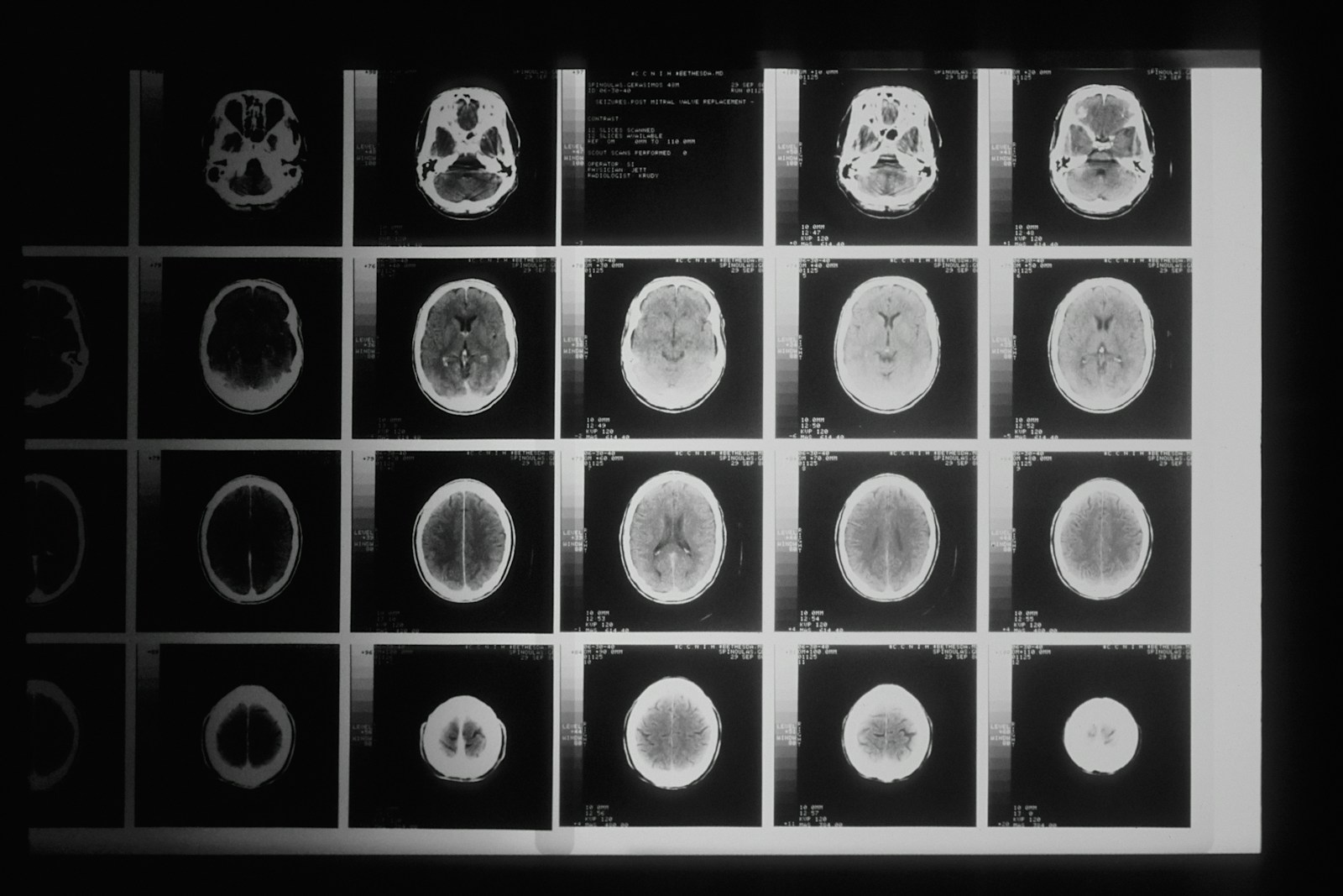
Each neuroimaging modality answers a specific biological question, so selection should follow the drug’s mechanism rather than whichever technique currently attracts attention. The imaging biomarkers available to CNS sponsors extend beyond PET tracers, while established biomarker programs continue to select participants and demonstrate target engagement. The next step is choosing a measure that reflects the process the therapy claims to alter.
DTI and White Matter Integrity
Diffusion tensor imaging, or DTI, is a form of MRI brain imaging that measures how water moves through tissue. In organized white matter, water tends to diffuse along nerve fibers. Fractional anisotropy describes this directional organization, while diffusivity captures the amount and pattern of movement.
Changes in these measures can reveal axonal injury or demyelination before the damage appears as gross tissue loss. This makes DTI relevant to traumatic brain injury (TBI), where conventional scans can miss diffuse injury, and vascular contributions to dementia, where small-vessel damage disrupts communication between brain regions.
However, DTI is sensitive to scanner settings, motion correction, gradient directions, and tract definitions. Consistent acquisition and processing are therefore necessary for meaningful comparisons.
Arterial Spin Labeling and Cerebral Perfusion
Arterial spin labeling, commonly called ASL, uses magnetically labeled blood water as an internal tracer to estimate cerebral blood flow. Because it does not require an injected contrast agent, it can be repeated across scheduled visits without adding contrast exposure.
ASL is most informative when a therapy is expected to affect hypoperfusion, vascular function, or neurovascular coupling. Regional blood-flow changes may provide an earlier view of biological activity than structural loss, which develops over a longer period.
Still, blood-flow measurements respond to motion, labeling efficiency, transit time, and physiological conditions around the scan. Protocols need consistent timing and acquisition procedures when ASL supports longitudinal comparisons.
Volume Loss Outside the Hippocampus
Hippocampal volume remains familiar in Alzheimer’s disease research, but regional volumetrics can track patterns that the hippocampus alone does not capture. Frontal and parietal cortical thickness, basal ganglia volumes, and brainstem measurements can align imaging analysis with Parkinson’s disease, frontotemporal presentations, and atypical phenotypes.
This regional approach supports precision medicine by testing whether change occurs in the anatomy implicated by a disease pattern or therapeutic mechanism. Whole-brain volume can dilute a localized effect, while a pre-specified regional measure provides clearer biological interpretation.
Neuroinflammatory imaging applies similar logic to glial activity, but it remains earlier in methodological maturity. Variability in tracer behavior, analysis, and repeatability currently limits its role in confirmatory endpoint sets.
Where Imaging Belongs in the Endpoint Hierarchy
The useful question is not simply whether a novel imaging measure is scientifically credible. Instead, sponsors must determine what job it performs in the clinical trial design. An endpoint used to demonstrate target engagement carries a different evidentiary burden from one intended to predict clinical benefit, determine dose, or support regulatory approval. Defining that role before data collection prevents an interesting scan result from being overinterpreted later.
Secondary Endpoints That Carry Real Weight
Novel imaging measures often do their best work as secondary or supportive endpoints. They can provide a mechanistic explanation for a clinical signal, distinguish biological inactivity from a poorly chosen dose, and deliver a shorter-horizon readout than many cognitive assessments.
Their value increases when they move in the same direction as the rest of the endpoint set. For example, a perfusion or white matter change that aligns with cognitive assessments and quality-of-life (QOL) endpoints creates a more coherent account than any measure in isolation.
Digital biomarkers add continuity between scanning visits by capturing behavior more frequently. Imaging provides anatomical or physiological depth, while digital measures show whether related changes appear in everyday function.
What Regulators Expect You to Validate
Primary endpoint status generally requires evidence that change in an imaging measure corresponds to meaningful clinical benefit. Most emerging measures remain secondary by design because that relationship has not been established strongly enough for the FDA to treat the scan as a substitute for patient-centered outcomes.
Endpoint selection and validation involve more than biological plausibility. Sponsors need to establish analytical validity, define how each image becomes a numerical result, and demonstrate reproducibility across scanners and visits.
The analysis plan must pre-specify acquisition procedures, quality thresholds, regions of interest, exclusions, and statistical handling. A documented context of use should also define the population, disease stage, and decision the measure supports. These conditions need agreement before the data exist, not after an encouraging pattern appears.
Making Multi-Site Imaging Data Worth Analyzing
Scanner variability is often the largest operational threat to a novel imaging endpoint. Differences in manufacturer, field strength, head coil, reconstruction software, and sequence version can create between-site variation that obscures the treatment-related change a study is designed to detect.
Harmonized acquisition protocols must be ready before the first participant is scanned. Phantom qualification checks whether each scanner produces measurements within defined limits, while locked sequence parameters prevent local adjustments during recruitment. Scanner maintenance or software upgrades should trigger documented review rather than pass unnoticed into the dataset.
Central reading reduces another source of inconsistency. For volumetrics and DTI, segmentation rules, tract definitions, motion correction, and other processing choices directly shape the reported number. Blinded independent analysis applies those choices consistently and prevents treatment assignment or visit order from influencing interpretation.
Site training and data-quality controls complete the process. Technologists need clear instructions for participant positioning, motion management, sequence selection, and scan transfer. Real-time review should flag incomplete coverage, artifacts, or protocol deviations while repeat imaging remains possible.
These controls affect drug development timelines because qualification, training, and central review add setup time and cost. The practical trade-off is to include fewer, better-standardized MRI brain imaging endpoints tied directly to the mechanism rather than a broad exploratory panel that produces inconsistent data.
What This Means for the Next CNS Protocol
The move beyond amyloid and tau is not a rejection of proteinopathy imaging. Instead, it widens what CNS clinical trials measure so that target engagement can be interpreted alongside the vascular, structural, and inflammatory processes involved in neurodegenerative diseases.
Endpoint choice should follow the drug mechanism and the biology the therapy claims to change. Novelty alone does not make a measure submission-ready. Standardized acquisition, reproducible analysis, and a defined place within the endpoint hierarchy determine whether the resulting data can support the clinical argument.