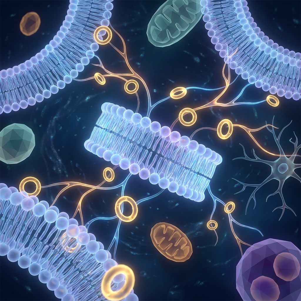
Plasmalogens Function: 6 Biological Roles Science Has Confirmed
Introduction: Why Plasmalogen Function Matters More Than You Think
Plasmalogens account for roughly 15 to 20% of all phospholipids in human cellular membranes, making them one of the most abundant lipid classes in the body. Yet most people have never heard of them.
Plasmalogens are a unique subclass of glycerophospholipids, distinguished by a vinyl-ether (plasmenyl) bond at the sn-1 position and a polyunsaturated fatty acid (PUFA) at the sn-2 position. That single structural distinction drives biology that no ordinary phospholipid can replicate.
Rather than narrowing the story to Alzheimer’s disease (the angle most articles fixate on), this piece presents six mechanistically distinct biological roles confirmed by science, spanning the brain, heart, immune system, and mitochondria. The momentum is real: as of 2025, more than 100 peer-reviewed publications per year are devoted to plasmalogen biology.
Two primary subtypes recur throughout: ethanolamine plasmalogens (PlsEtn) and choline plasmalogens (PlsCho). Their tissue-specific roles matter, and this article draws on research through 2026, including a landmark Biophysical Journal study and a ScienceDirect mitochondrial fission study, to provide the most current picture available.
What Makes Plasmalogens Structurally Unique
The defining feature is the vinyl-ether (plasmenyl) bond at the sn-1 position. Conventional phospholipids carry an ester bond in that same spot; plasmalogens do not.
On the glycerol backbone, the sn-1 position carries the vinyl-ether-linked fatty alcohol, the sn-2 position carries a PUFA (commonly DHA, EPA, or arachidonic acid), and the sn-3 position carries the phosphate head group. That head group comes in two variants: ethanolamine (forming PlsEtn) and choline (forming PlsCho).
Tissue abundance grounds the significance. Up to 70% of ethanolamine glycerophospholipids in myelin are plasmalogens, roughly 30 to 40% of choline glycerophospholipids in the heart are plasmalogens, and about 20% of brain glycerophospholipids are plasmalogens. Ethanolamine plasmalogens dominate in the brain, neurons, and mitochondria, while choline plasmalogens are especially abundant in the heart and cardiovascular tissues. The vinyl-ether bond alters phospholipid packing density, membrane curvature, and chemical reactivity in ways ester-linked lipids simply cannot match.
The Peroxisome–Plasmalogen Biosynthesis Axis: The Upstream Story Competitors Miss
Plasmalogen biosynthesis is a two-compartment process. The rate-limiting steps occur in peroxisomes, and the pathway is completed in the endoplasmic reticulum. Two exclusively peroxisomal enzymes, GNPAT and AGPS, initiate vinyl-ether bond formation.
The clinical proof of concept is stark. Peroxisomal biogenesis disorders such as Zellweger spectrum disorder and rhizomelic chondrodysplasia punctata (RCDP) cause severely reduced plasmalogen levels, and affected individuals show reduced lifespans, abnormal neurological development, skeletal dysplasia, impaired respiration, and cataracts.
This axis also explains aging. Plasmalogen levels decline with age because of reduced peroxisome biogenesis, impaired peroxisomal protein import, and increased activity of the degrading enzyme PlsPLA2. A 2026 study confirmed age-related decline in peroxisome biogenesis in human B cells. Most competing content describes plasmalogen decline as a consequence of aging without explaining that declining peroxisome function is the upstream cause. Because biosynthesis is peroxisome-dependent, circulating plasmalogen levels serve as measurable biomarkers of peroxisomal health, and ARUP Laboratories now offers a clinical test for red blood cell plasmalogens.
Role 1: Membrane Architecture and Dynamics
Plasmalogens are not passive components. They actively regulate membrane physical properties. The vinyl-ether linkage alters packing density and conformational order, influencing membrane thickness, fluidity, and lateral pressure profiles.
They also modulate membrane curvature, a property essential for vesicle formation, membrane fusion, and fission. In neurons, these events underpin synaptic vesicle cycling, neurotransmitter release, and receptor trafficking. Plasmalogens are further required for lipid raft integrity, the cholesterol- and sphingolipid-enriched microdomains that host integral membrane proteins. PlsEtn is central to neuronal and mitochondrial architecture, while PlsCho supports cardiomyocyte membranes, as detailed in a 2021 Frontiers in Cell and Developmental Biology review.
Role 2: Endogenous Antioxidant Defense
The vinyl-ether bond is preferentially targeted by reactive oxygen species (ROS), including peroxyl radicals, singlet oxygen, metal ions, and halogenating species. When ROS attacks that double bond, the plasmalogen is oxidized and destroyed; in the process, it neutralizes the oxidant and prevents chain-reaction damage to neighboring PUFAs and lipoproteins.
Because DHA and similar PUFAs are highly susceptible to oxidative damage, a plasmalogen shield in the same membrane is a critical protective strategy, especially in the oxidatively stressed brain. As plasmalogen levels fall, membrane antioxidant capacity drops, accelerating damage in a vicious cycle. A seminal 2004 review established this sacrificial mechanism. Unlike circulating free-radical scavengers, plasmalogens are membrane-integrated antioxidants operating precisely at the site of attack.
Role 3: PUFA Reservoir and Bioactive Lipid Precursor
Plasmalogens are a primary storage pool for DHA, EPA, and arachidonic acid, esterified at the sn-2 position. Free PUFAs cannot be stored without toxicity, so membrane integration provides a stable reservoir.
Phospholipase A2 enzymes cleave the sn-2 PUFA on demand. Released arachidonic acid becomes prostaglandins, thromboxanes, and leukotrienes that regulate inflammation, platelet aggregation, and vascular tone. DHA released from PlsEtn supports neuronal membrane fluidity and serves as a precursor for neuroprotective docosanoids. A 2022 clinical trial showed that oral DHA-AAG plasmalogen precursors dose-dependently increased blood DHA plasmalogen levels, confirming this reservoir is therapeutically targetable.
Role 4: Cell Signaling and Signal Transduction
Plasmalogens actively participate in signaling cascades. They maintain the lipid rafts that concentrate signaling receptors, regulate protein kinase C delta (PKCδ) activity involved in cell survival and apoptosis, and modulate ion channels that influence electrical excitability and synaptic transmission.
They also regulate cell adhesion molecules essential for immune cell recruitment and tissue repair, and they modulate Toll-like receptor 4 (TLR4) endocytosis, influencing inflammatory thresholds. Plasmalogen-derived lysophospholipids and fatty aldehydes act as second messengers, propagating signals from membrane to intracellular effectors.
Role 5: Anti-Inflammatory Regulation
Plasmalogens play a nuanced, bidirectional role in inflammation, regulating the balance between pro- and anti-inflammatory states. They inhibit TLR4 endocytosis, dampening innate immune activation. Yet they also serve as precursors for platelet-activating factor (PAF), a potent pro-inflammatory mediator, meaning plasmalogen levels influence PAF availability. Their arachidonic acid reservoir likewise governs the magnitude of eicosanoid-driven inflammation.
As levels fall with age, this regulatory capacity weakens, contributing to the chronic low-grade “inflammaging” of later life. A 2025 Cell Reports Medicine study of 187 human carotid plaques found a positive correlation between ether lipid phospholipids and more stable atherosclerotic plaque profiles, reinforcing the cardiovascular relevance of PlsCho.
Role 6: Neuronal Excitability, Synaptic Function, and BDNF Regulation
A 2026 Biophysical Journal finding demonstrated that plasmalogen-dependent modulation of ordered membrane domains directly controls neuronal excitability. By shaping lipid rafts at synapses, plasmalogens regulate receptor clustering and ion channel function; by enhancing membrane flexibility, they support vesicle fusion and neurotransmitter release.
They also regulate brain-derived neurotrophic factor (BDNF), a master regulator of synaptic plasticity and memory. Because PlsEtn dominates in neurons and myelin, its decline has outsized consequences. Ethanolamine plasmalogen levels are significantly reduced in Alzheimer’s disease brains, with dementia severity correlating with cortical and hippocampal plasmalogen content. Reduced levels also appear in Parkinson’s disease, ALS, and multiple sclerosis, and circulating PlsEtn indices correlate with cognition scores and CSF tau, as summarized in a 2026 BRMI article.
The 2026 Frontier: Plasmalogen Decline and Mitochondrial Fission Dynamics
The most recent mechanistic breakthrough connects plasmalogens to mitochondrial quality control. A 2026 ScienceDirect study in Drosophila found that mitochondrial-specific plasmalogen content decreases with age, and that plasmalogen biosynthesis mutants abolish stress-induced mitochondrial fission and Drp1 localization to mitochondria.
Fission segregates damaged components for removal via mitophagy. When fission fails, damaged mitochondria accumulate. Drp1 recruitment to the outer membrane appears to depend on plasmalogen-derived membrane properties, establishing a direct mechanistic link. As peroxisome function declines, plasmalogen biosynthesis falls, mitochondrial content drops, fission stalls, and energy production deteriorates. Since PlsEtn dominates mitochondrial membranes, this is a specifically PlsEtn-dependent phenomenon, complemented by a 2026 PMC review on peroxisomes in aging.
Plasmalogen Decline With Age: What the Research Shows
Levels fall progressively after midlife through three converging mechanisms: reduced peroxisome biogenesis, impaired protein import, and increased PlsPLA2 degradation. Disease-associated declines appear in Alzheimer’s, Parkinson’s, ALS, multiple sclerosis, metabolic syndrome, and type-2 diabetes, establishing this as a multi-disease phenomenon. Higher levels correlate with lower dementia risk and increased lifespan, including in beautiful aging centenarian research. In the heart, PlsCho decline is tied to less stable plaques. Because levels are measurable in blood, they serve as actionable biomarkers of biological aging, as reviewed in a 2020 Advances in Gerontology paper.
Plasmalogen Replacement Therapy: The Emerging Science of Restoration
Plasmalogen Replacement Therapy (PRT) aims to restore levels across neurodegenerative, cardiovascular, peroxisomal, and metabolic conditions. The Goodenowe et al. 2022 trial in 22 cognitively impaired persons showed oral DHA-AAG precursors dose-dependently raised blood DHA plasmalogens, reduced MDA, increased catalase activity, and were associated with cognition and mobility improvements over four months.
Because plasmalogens degrade in the gut, effective supplementation requires alkylglycerol-based precursors that bypass degradation and convert in target tissues. A Phase 1 trial of precursor PPI-1011 (2023) advanced this approach for RCDP, and a 2025 FASEB BioAdvances review highlighted nanomedicine delivery to overcome bioavailability challenges. Not all products deliver meaningful precursor concentrations. Prodrome Science applies the precursor strategy directly, offering 900 mg per serving versus other products on the market at 0.5 to 4 mg per capsule. These statements have not been evaluated by the Food and Drug Administration. This product is not intended to diagnose, treat, cure, or prevent any disease.
Conclusion: The Full Biological Picture of Plasmalogen Function
Six confirmed roles emerge: membrane architecture, antioxidant defense, PUFA reservoir, cell signaling, anti-inflammatory regulation, and neuronal excitability. The 2026 mitochondrial fission finding adds an emerging seventh dimension. The PlsEtn versus PlsCho distinction remains a key takeaway: ethanolamine plasmalogens govern brain and mitochondrial function, while choline plasmalogens protect the cardiovascular system. Declining peroxisome function, not aging itself, is the root cause of plasmalogen loss. With more than 100 publications per year, plasmalogen science is advancing rapidly, and measuring and supporting these levels is now a practical, evidence-based option.
Take the Next Step With Prodrome Science
Prodrome Science was built on 30-plus years of lipid and metabolomic research focused specifically on plasmalogen biology, the science covered throughout this article.
For readers curious about their own status, the ProdromeScan™ blood test measures over 40 biomarkers, including plasmalogen levels, providing a personalized biochemical baseline. Note that the ProdromeScan is available to qualified health professionals; consumers should contact Prodrome Science directly to inquire about eligibility. The precursor product line aligns with the distinctions above: ProdromeNeuro™/PlasmalogenN3™ targets gray matter and brain (Omega-3 PlsEtn), while ProdromeGlia™ targets white matter (Omega-9). The premium PC+ line (Glia PC+ and Neuro PC+) offers higher-concentration support. Health practitioners can access wholesale pricing, clinical tools, and the Elite Practitioner training program through Prodrome’s professional-first platform.
Explore the ProdromeScan test, browse the plasmalogen line, or contact Prodrome Science at cs@prodrome.com. Supporting plasmalogen levels before illness takes root is the biochemical prodrome approach to lasting health.
These statements have not been evaluated by the Food and Drug Administration. This product is not intended to diagnose, treat, cure, or prevent any disease.